

An innovative artificial intelligence (AI) tool is transforming the training of medical imaging software, allowing doctors and researchers to easily and cost-effectively complete training tasks with only a small amount of patient scan data. This tool revolutionizes the medical image segmentation process, which aims to label each pixel in an image according to its meaning, such as distinguishing cancerous tissue from normal tissue. Traditionally, this task has relied on highly trained experts, and although deep learning has shown potential for automation, it has an urgent need for large volumes of pixel-by-pixel annotated image data.
Dr. Li Zhang and her research team at the University of California San Diego developed this new AI tool to address the challenge. Professor Zhang pointed out: "Deep learning requires massive amounts of data, but in many medical scenarios, such datasets simply do not exist." The new tool can learn image segmentation from just a small number of expert-labeled samples, reducing data requirements by up to 20 times and providing faster and more economical diagnostic tools for hospitals and clinics with limited resources.
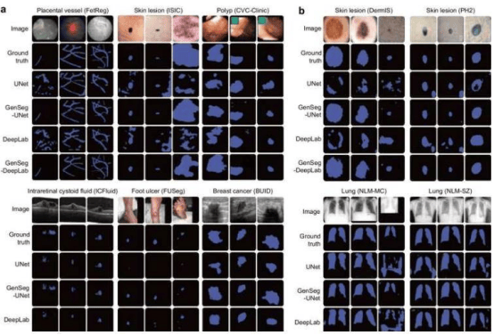
The research results were published in the journal Nature Communications. The AI tool has been validated in various medical image segmentation tasks, including the identification of skin lesions, breast cancer, placental vessels, colon polyps, and foot ulcers. It can also handle 3D images, such as the delineation of the hippocampus or liver. In data-scarce environments, the tool outperforms existing methods by 10% to 20%, reduces the amount of real-world training data required by 8 to 20 times, and often delivers superior performance.
Professor Zhang used dermatological diagnosis as an example to illustrate how the tool assists doctors. Traditionally, experts need to annotate thousands of images, whereas the new tool requires only 40 images to identify suspicious lesions in dermoscopy images in real time. "It helps doctors make diagnoses faster and more accurately," Professor Zhang said. The system operates in stages, augmenting small datasets by generating synthetic image masks and using a feedback loop to optimize image generation, ensuring that the synthetic data is both realistic and improves the model's segmentation capability.