en.Wedoany.com Reported - A team from Nanyang Technological University, Singapore (NTU Singapore) has developed an imaging strategy that requires no exogenous labels, utilizing wide-field interferometric scattering (iSCAT) microscopy to map subcellular dynamics in living cells. Published in PhotoniX Life, this study reveals cell states and dynamic heterogeneity without adding labels through statistical analysis of random fluctuations in iSCAT image sequences.
As dynamic systems, cells exhibit thermal and active motions in their organelles, macromolecules, membranes, and cytoskeletal structures, which are related to subcellular architecture and energy states. Directly visualizing these rapid and spatially heterogeneous motions in living cells is challenging, and common exogenous labels may interfere with cellular behavior.
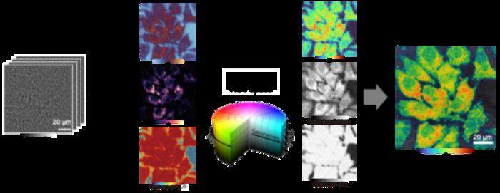
To address this challenge, the research team performed a full-field analysis of the power spectral density (PSD) of high-speed iSCAT time series. Across multiple cell types, the PSD of iSCAT signals in most cellular regions followed an inverse power-law relationship in the 30–1250 Hz range: S(f)=βf⁻α. The fitted spectral exponent (α) and amplitude (β) reflect the characteristics and intensity of subcellular motion, respectively. By encoding α as hue, β as brightness, and goodness of fit (R²) as saturation, two-dimensional spectral exponent maps were generated in hue-saturation-value (HSV) space to reflect the spatial distribution of cellular dynamics.
Experimental results show that this method enables label-free visualization of changes in cell state. In HeLa cells, spectral exponent maps clearly distinguished mitotic from interphase cells and tracked dynamic transitions during mitosis. In hydrogen peroxide-induced apoptotic cells, the maps revealed increased heterogeneity, with high-α regions associated with apoptosis-related membrane blebbing. The maps also captured differences related to malignancy in thyroid cancer cell subtypes: papillary thyroid carcinoma (PTC), follicular thyroid carcinoma (FTC), and anaplastic thyroid carcinoma (ATC) cells showed a gradual decrease in median spectral exponent values with increasing malignancy.
The findings indicate that wide-field iSCAT imaging combined with power spectral exponent analysis can serve as an intrinsic optical readout of cell state. Due to the absence of exogenous labels, this method may have potential applications in longitudinal live-cell studies, mechanobiology, cancer research, and quality assessment in stem cell therapy.
See the article: “Label-free mapping of subcellular dynamics using wide-field interferometric scattering microscopy and spectral exponent analysis.” PhotoniX Life. doi: 10.3724/PXLIFE.2025-0012
This article is compiled by Wedoany. All AI citations must indicate the source as "Wedoany". If there is any infringement or other issues, please notify us promptly, and we will modify or delete it accordingly. Email: news@wedoany.com