en.Wedoany.com Reported - A collaborative team in France has developed Otosurg, a multi-material 3D-printed ear surgery simulator designed specifically for surgical training in transcanal ear procedures. The device combines clinical realism, anatomical customization, and validated competency assessment.
The project was completed by Mael Duportal, Additive Manufacturing and CAD Engineer at M3DPrint; Juliette Prebot, Chief R&D Engineer at PRIM3D, AP-HP (Greater Paris University Hospitals); and François Simon, Consultant ENT Surgeon at AP-HP and Professor at Université Paris Cité. The project aims to address the clinical challenge of training surgeons to perform procedures they have never executed without putting patients at risk.
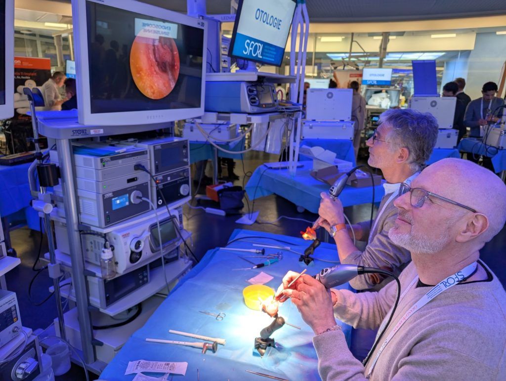
Traditional surgical training tools each have limitations: human cadavers pose ethical constraints and cannot replicate pathology, animal models differ anatomically from humans, virtual reality lacks credible haptic feedback, and catalog-based simulators cannot adapt to specific diseases or personalized learning needs. Simon noted that surgical training is not a single threshold to cross, but a series of "firsts": the first time performing a surgery, the first time without a mentor present, and the first time encountering complications. Otosurg targets this gap. The technique uses an endoscope inserted directly into the ear canal rather than making an incision behind the ear. The shift from microscope to endoscope represents an entirely new set of skills.
Otosurg serves two user groups simultaneously: residents encountering otologic surgery for the first time, and experienced surgeons with decades of practice who need to retrain from microscope to endoscopic techniques. Simon explained that trainees can perform at least six to eight procedures per day, which is not possible with cadaver models. The logic is progressive: the first full day is spent on the simulator handling different pathologies and disease variants, followed by cadaveric practice the next day.
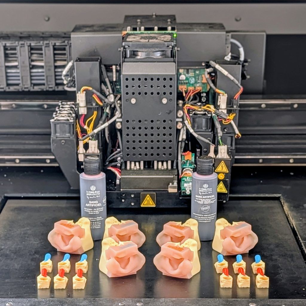
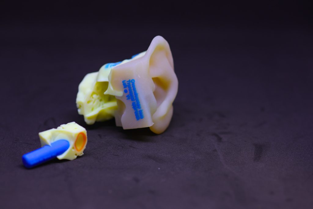
Prebot described the development process as methodical and iterative. The design phase utilized open-source tools: 3D Slicer for segmenting CT scan data into anatomical structures, and Blender for adapting these structures to manufacturing constraints. The ossicles are extremely small in reality and had to be slightly enlarged for printing. The model was built as a modular system: a reusable base and interchangeable cartridges, undergoing at least five complete design iterations before reaching the validated version, with a total development time of approximately one year. The final model uses Stratasys' PolyJet printing technology in anatomically critical areas, capable of combining multiple materials in a single build to replicate hard and soft tissues. The simulator also supports adding blood for dramatic effect during simulation, altering textures, introducing tissue adhesion, and recreating the visual complexity of a bleeding surgical field. Cartridges can be printed with non-physiological colors to guide beginners, or specific elements such as the tympanic membrane can be removed to isolate particular stages of surgery for focused training.
The team conducted a formal validation study with a group of experts and students, with results published in the peer-reviewed journal Otology & Neurotology. The training framework built around Otosurg incorporates the Objective Structured Assessment of Technical Skills (OSATS), a validated tool for stepwise evaluation of surgical competency. The simulator is currently part of a hybrid training curriculum developed in collaboration with the University of Toronto and is commercially distributed through M3DPrint in Europe, Canada, and the United States. The model is continuously refined based on user feedback from surgeons and institutions.
When asked what they would like to see from technology providers, the M3DPrint team pointed to openness issues. Current high-end systems like PolyJet are mostly closed ecosystems—proprietary software, proprietary materials—limiting the range of tissue simulations that can be developed. Duportal cited the Digital Anatomy Processor feature as a step in the right direction, allowing customized material blending, but called for greater freedom in software platforms for developers to design beyond predefined parameters. The cost gap between accessible technologies like FDM and high-fidelity multi-material systems remains a barrier. The team believes that the hardware to produce clinically meaningful surgical simulators already exists; the limiting factors lie in software flexibility, material openness, and the sustained clinical-engineering partnerships needed to convert anatomical data into training tools. Future directions for the project include customizable catalogs allowing institutions to order cartridges for specific pathologies or training objectives, as well as extending the same multi-material, clinically validated approach to other surgical specialties.
This article is compiled by Wedoany. All AI citations must indicate the source as "Wedoany". If there is any infringement or other issues, please notify us promptly, and we will modify or delete it accordingly. Email: news@wedoany.com