
en.Wedoany.com Reported - Researchers at the Centre for Addiction and Mental Health, University of Toronto, reported at the 2026 Annual Meeting of the Society of Nuclear Medicine and Molecular Imaging that SV2A PET imaging can detect reduced synaptic density in the spinal cord and brain of living multiple sclerosis patients and animal models. This study directly confirms that synaptic loss is a universal feature of multiple sclerosis, providing a new quantitative tool for disease monitoring and treatment evaluation.
Multiple sclerosis has traditionally been viewed as a disease that damages the protective layer of nerves, but there is another, more subtle form of injury: synaptic loss. Synapses are critical connection points where brain cells communicate. Although the spinal cord is a primary and often early site of inflammation and neuropathology in multiple sclerosis, in vivo quantification of synaptic density in this region had not been previously explored.
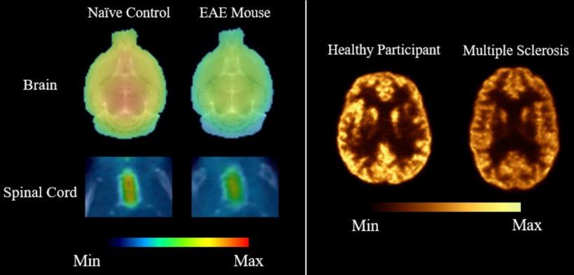
To fill this research gap, the team used a specialized imaging technique called SV2A PET to visualize and quantify connection loss in the spinal cord of living multiple sclerosis mouse models and patients. Researchers performed 18F-SynVesT-1 PET scans on mice with experimental autoimmune encephalomyelitis and healthy control mice, defined spinal cord regions of interest, and quantified and compared total distribution volume and radiotracer binding across groups. In collaboration with Yale University, the study also conducted 11C-UCB-J PET imaging on six multiple sclerosis patients and six healthy controls, generating total distribution volume maps and comparing human PET data.
In multiple sclerosis model mice, 18F-SynVesT-1 PET successfully detected significant reductions in synaptic density within specific spinal cord regions, confirmed by binding studies. In human PET studies, compared to healthy controls, multiple sclerosis patients showed a 16.4% reduction in 11C-UCB-J binding across the whole brain, with widespread reductions also observed in subcortical and spinal cord regions. Dr. Chao Zheng from the Centre for Addiction and Mental Health, University of Toronto, noted that this work establishes a translational framework linking preclinical findings with preliminary human PET imaging studies. According to lead presenter Pou Hong Justin Chia, understanding where and how these connections are lost can help explain patient symptoms and provide clinicians and researchers with more sensitive methods to detect disease-related changes and monitor progression.
Currently, SV2A PET imaging in multiple sclerosis can be used in clinical trials at specialized academic centers. The data from this study represent a necessary step toward larger-scale clinical trials; if validated in larger studies, this imaging method could potentially be integrated into clinical practice and drug development in the future.
Abstract 261222. "Tracking Synaptic Density Loss in the Spinal Cord of Mice with Experimental Autoimmune Encephalomyelitis and Preliminary Evaluation of SV2A PET in Multiple Sclerosis Patients," Pou Hong Justin Chia, Hannah Le, Junchao Tong, Mohammad Alijaniaram, Neil Vasdev, and Chao Zheng, Centre for Addiction and Mental Health (CAMH), University of Toronto; and Takuya Toyonaga and Ming-Kai Chen, Yale University, New Haven, Connecticut.
This article is compiled by Wedoany. All AI citations must indicate the source as "Wedoany". If there is any infringement or other issues, please notify us promptly, and we will modify or delete it accordingly. Email: news@wedoany.com