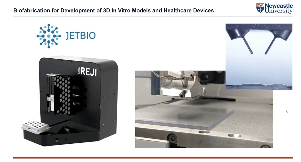
en.Wedoany.com Reported - Dr. Priscila Melo, a Lecturer in Bioengineering at Newcastle University and co-founder of JetBio, is leading a team to construct 3D tissue models using their developed Reactive Jet Impingement (ReJI) bioprinting technology, aiming to transform how new drugs are evaluated before entering clinical application.
Current preclinical drug screening primarily relies on 2D cell culture, where cells grow as a monolayer on a flat substrate. Approximately 75% of new drugs entering Phase I clinical trials ultimately fail, most commonly due to insufficient efficacy or safety issues that were not detected in early testing. Human cells exist within a three-dimensional extracellular matrix that not only regulates the mechanical environment but also influences nutrient diffusion, intercellular signaling, and tissue-specific functions—complexity that 2D models cannot replicate. Regulatory trends are also driving this shift. In 2023, the U.S. Food and Drug Administration (FDA) stated that validated in vitro models could serve as the basis for human drug trials without requiring animal testing. Europe has since aligned with this direction, and the UK is working to implement similar standards for specific test categories by 2030.
Dr. Melo stated that 3D printing offers a more accurate and viable alternative, aiming to eliminate or reduce animal testing as much as possible. At Newcastle University, her team's ReJI technology deposits droplets simultaneously from two cartridges via microvalves—one containing a hydrogel precursor and the other carrying a cell suspension with a crosslinking agent. The droplets interact in mid-air, producing structured cell constructs within milliseconds. This platform has demonstrated compatibility with substrates such as synthetic fibers, metals, and biological tissues.
In cardiotoxicity screening, the team used a bioink containing type I collagen, alginate, and fibrinogen to print a cardiac tissue model comprising HL-1 cardiomyocytes at a density of 5 million cells per milliliter of gel—the highest reported density for this cell type. The resulting constructs maintained spontaneous contractile activity for up to 21 days, whereas the same cells lost beating function within approximately 7 days in standard 2D culture. Electrical activity assessed via multielectrode arrays became progressively more organized over time, and the model responded appropriately to both proarrhythmic and antiarrhythmic drugs.
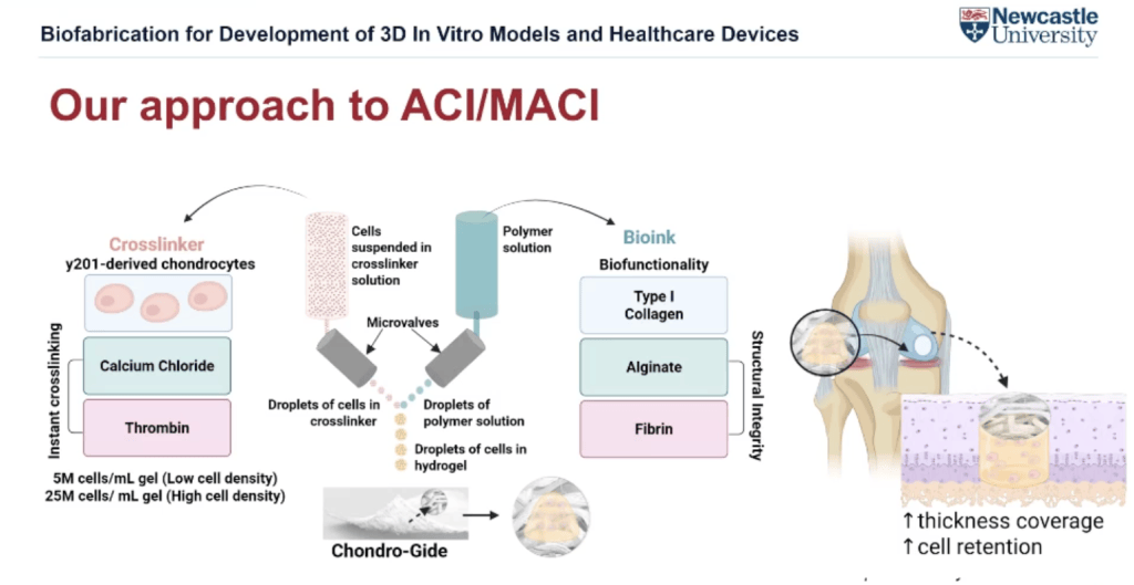
In cartilage repair studies, the team directly bioprinted chondrocyte-laden hydrogel onto Chondro-Gide (a clinically available collagen-based repair patch), achieving superior cell distribution and cartilage-specific marker expression compared to all other tested conditions. The results indicate that integrating bioprinted constructs with existing scaffold materials can significantly enhance the biological performance of current cartilage therapies.
Dr. Melo's work is part of an industry trend toward 3D bioprinted tissue models that more faithfully replicate the in vivo environment. The ultimate goal is to develop multi-physiological systems capable of reproducing systemic interactions, enabling connections between different tissues and modeling of comorbidities. CELLINK has listed reducing and eliminating animal testing as a commercial priority; the EU-funded BRIGHTER project, coordinated by the Institute of Bioengineering of Catalonia, is developing bioprinting processes aimed at reducing reliance on animal models in tissue engineering and regenerative medicine. Additionally, researchers at TU Wien have used multiphoton lithography to fabricate human tissue-on-a-chip constructs, and a bioprinted cerebrovascular model capable of replicating atherosclerotic flow conditions has also been developed, demonstrating the feasibility of recreating increasingly complex physiological environments in vitro.
This article is compiled by Wedoany. All AI citations must indicate the source as "Wedoany". If there is any infringement or other issues, please notify us promptly, and we will modify or delete it accordingly. Email: news@wedoany.com