
en.Wedoany.com Reported - Researchers from University College London (UCL) and the European Synchrotron Radiation Facility (ESRF) have, for the first time, created a three-dimensional map of the cardiac conduction system in patients with Tetralogy of Fallot using Hierarchical Phase-Contrast Tomography (HiP-CT). The map reveals anatomical features that may lead to cardiac conduction disorders in patients, providing clearer navigation for surgeons. The study, part of the international Human Organ Atlas collaboration, was published in The Journal of Thoracic and Cardiovascular Surgery.
Congenital heart disease affects approximately 1% of the global population. Many infants require heart surgery after birth, and while survival rates are high, some patients may later develop complications such as arrhythmias. Surgeons have long faced a challenge: the heart's delicate conduction system is invisible during surgery, and any disruption can cause problems. Professor Andrew Cook, a cardiac anatomy expert at UCL and senior author of the study, likened this dilemma to home renovation: "You wouldn't start drilling holes in a wall without knowing where the wires are. The same principle applies to the heart."
The HiP-CT technology used by the research team was developed by an international team led by UCL at the ESRF in Grenoble, France. This technology utilizes a new generation of synchrotron radiation sources, providing X-ray beam intensities millions of times higher than traditional hospital CT scanners. It can non-destructively scan entire human organs ex vivo and magnify them to near-cellular resolution, down to 2 micrometers. Dr. Joseph Brunet, a UCL researcher and visiting scientist at the ESRF, explained that radiology and histology have provided very different views of the body for over a century, and HiP-CT technology ultimately bridges this gap.
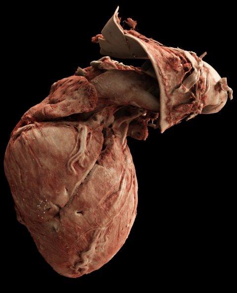
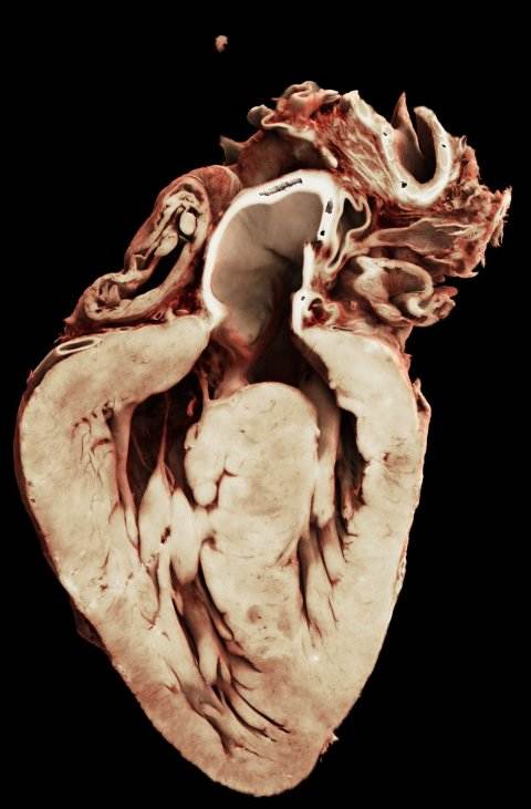
Using HiP-CT technology, the researchers non-destructively examined 18 complete human heart specimens, both diseased and healthy. Through 3D reconstruction of the scan data, the team mapped the fine fiber network of the heart's conduction system. The results showed that in patients with Tetralogy of Fallot, the electrical conduction pathways in the right ventricle are thinner than in healthy hearts and are distributed on the ventricular septum, resembling a cloth covering the surface. This finding provides surgeons with a clearer image of the anatomical structure. Mr. Adrian Crucean, a specialist congenital cardiac surgeon at Birmingham Children's Hospital and Queen Elizabeth Hospital in the UK, stated that any new knowledge about the heart's anatomy can help improve surgical techniques in a highly challenging environment.
Dr. Monique Jongbloed, an adult congenital heart disease specialist at Leiden University Medical Center and a member of the EuReCCA consortium, noted that many adult patients who underwent successful surgery in childhood later developed complications such as arrhythmias. "This information is undoubtedly a game changer, reshaping our understanding of the structure and precise anatomical location of the cardiac conduction system in congenital heart disease."
The researchers also developed computational tools capable of analyzing and visualizing data in an immersive 3D environment. Dr. Vaishnavi Sabarigirivasan, a UCL PhD student and corresponding author of the paper, explained that this information can be brought into virtual reality and 3D printed, and can be used to train surgeons. It has never before been possible to see the conduction system in this way. Dr. Paul Tafforeau, an ESRF scientist and pioneer of imaging technology for the Human Organ Atlas, stated that HiP-CT technology was initially developed during the COVID-19 pandemic to study human lungs. Within a few years, data quality and acquisition speed have dramatically improved, enabling the scanning of enough organs for relevant pathological studies. Professor Peter Lee, Chair of Mechanical Engineering at UCL and Principal Investigator of the HOA beamline, emphasized that the Human Organ Atlas brings together scientists and doctors from nine institutions worldwide and is continuously expanding, helping to gain new insights into diseases from osteoarthritis to heart disease. Dr. Claire Walsh, Associate Professor of Mechanical Engineering at UCL and Director of the Human Organ Atlas Hub, pointed out that the atlas showcases team science at its best and is an incredible resource that will continue to grow.
Members of the EuReCCA consortium, from London, Paris, Leiden, and Birmingham, possess specific expertise in cardiac structural architecture and are extending their research to other forms of congenital heart disease, such as "single ventricle disease." Their goals include conducting "deep phenotyping" of congenital and acquired heart diseases, rapidly translating research into clinical practice and surgical training, and generating open-access data to support scientists in creating "digital twins" of heart function.
This article is compiled by Wedoany. All AI citations must indicate the source as "Wedoany". If there is any infringement or other issues, please notify us promptly, and we will modify or delete it accordingly. Email: news@wedoany.com