en.Wedoany.com Reported - Researchers at Washington University in St. Louis have developed a portable bedside positron emission tomography (PET) system capable of real-time imaging of any organ, providing high-quality guidance for interventional procedures such as biopsies and tumor ablation. The study was presented at the 2026 Annual Meeting of the Society of Nuclear Medicine and Molecular Imaging.
Current interventional procedures primarily rely on anatomical imaging modalities such as ultrasound, X-ray fluoroscopy, and CT for guidance. Studies have shown that dedicated PET/CT-guided interventional radiology procedures offer higher accuracy, but their high cost limits widespread adoption in most hospitals.
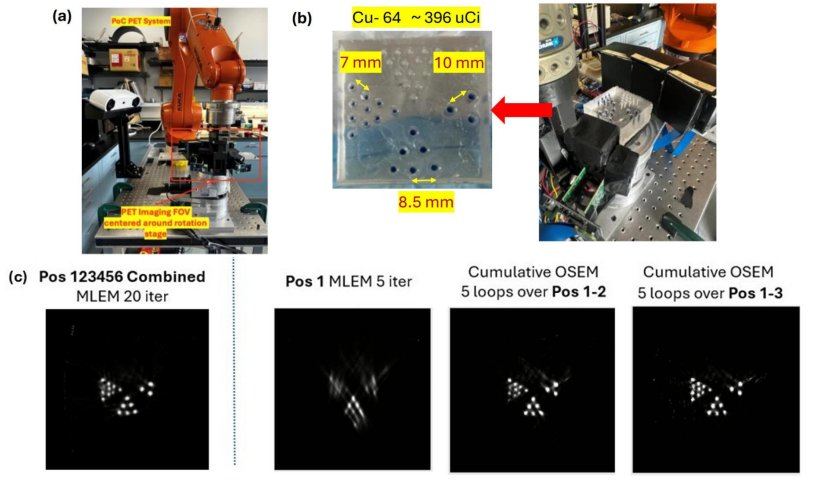
Dr. Yuan-Chuan Tai, senior author of the study and a researcher at Washington University in St. Louis, stated that a portable PET device with real-time imaging capabilities could bring molecular imaging information into interventional procedures. To this end, the team developed a portable bedside PET system equipped with a robotic arm, allowing the detector panel to be positioned anywhere to image any organ of interest. The study used this portable system to explore the feasibility of interactive PET scanning and real-time image update strategies. During phantom imaging of a phantom containing three sets of radioactive tracer-filled rods, the detector panel was moved to six user-selected positions. Image reconstruction began with five iterations at the first position, followed by alternating single-iteration updates as data from each new position was acquired. Since data acquisition time was significantly longer than reconstruction time, images were continuously updated during the acquisition process. Additionally, the study compared results using a traditional PET reconstruction framework, where images are generated after scanning is complete.
The image quality of the portable bedside PET system using the real-time image update framework was comparable to that of the traditional reconstruction framework. Phantom structures became clearly distinguishable after scanning three to four positions, indicating that scanning could be terminated early if the imaging task was completed; alternatively, image quality could be further improved by increasing the number of scanning positions or reconstruction iterations.
Xiyan Li, a graduate research fellow in the Imaging Science PhD program at Washington University in St. Louis, noted that this approach better supports bedside interactive and adaptive imaging workflows, representing a paradigm shift and opening new avenues for deploying novel molecular imaging applications.
The current study used a benchtop prototype system. Researchers are building a prototype suitable for preliminary human imaging studies, which are expected to begin in 2027.
This article is compiled by Wedoany. All AI citations must indicate the source as "Wedoany". If there is any infringement or other issues, please notify us promptly, and we will modify or delete it accordingly. Email: news@wedoany.com